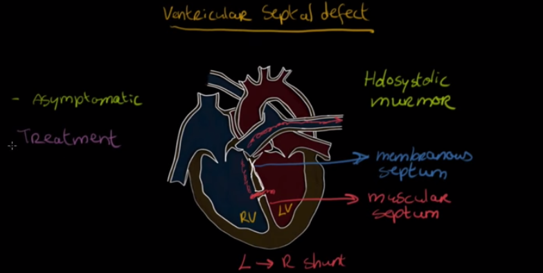
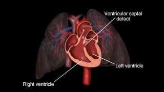
Postinfarction ventricular septal defect, or VSD, is an uncommon — but serious — complication of acute myocardial infarction and it’s often deadly.
Doctors say the event happens within 2-8 days after an infarction and often causes what’s known as cardiogenic shock. The condition requires that patients undergo emergency surgical treatment and coronary artery bypass grafting is often required.
The preferred treatment is known as percutaneous closure , and surgeons say it’s perhaps the most viable treatment strategy. In surgery, the word ‘percutaneous’ refers to any medical procedure intervention done via needle-puncture of the skin rather than by using a scalpel or laser. This percutaneous approach is often used in vascular procedures like angioplasty and stenting, and it uses a needle catheter and a wire placed into a blood vessel.
The Bristol Heart Institute, a leading center for the treatment of the condition using percutaneous intervention in their catheter lab, say using a physical representation of the defects will prove key in treating the damage.
Evan Ansell, Ian Negus, Margaret Saunders, Mark Turner, Nathan Manghat, and Mark Hamilton at the School of Cellular and Molecular Medicine of the University of Bristol and University Hospitals Bristol say a study they took proved that a 3D printer is ideal to reproduce representations of these ventricular septal defects.
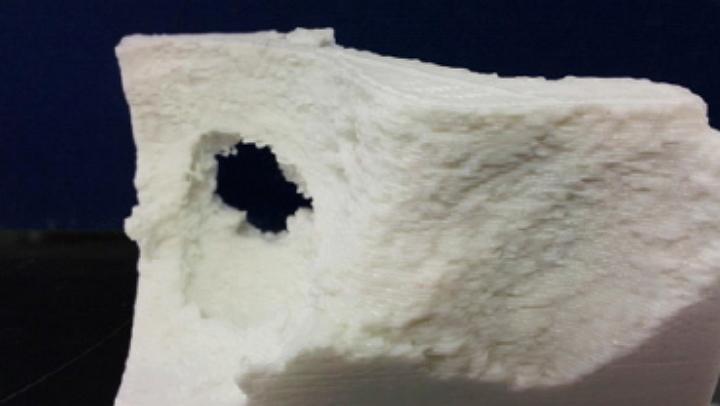
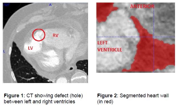
Researchers used a RepRap Mendel printer to build models segmented from CT scans to develop a method to take image data from what’s called CT Coronary Angiography. The data is used to create a 3D print of a VSD, and doctors can then use the models to assess the accuracy of the process and identify any limitations which might exist.
The team says the data was anonymized before software was used to select the area affected by the defect and then create a ‘segmented’ portion of the ventricular wall for review.
Doctors and researchers are now bring the full power of 3D printing technology to bear in their efforts to plan for delicate surgical procedures. Do you know of any ways medical professionals are using 3D printing to improve patient outcomes? Let us know in the 3D Printed Models to Save Lives forum thread on 3DPB.com.