Trepanning was once the front line treatment for various ailments from seizures to skull fractures to brain disorders. It was pretty grisly stuff, but the work of scientists and medical professionals with 3D printing is now making current facial and brain surgery techniques safer, and better planned, than the old “drill a hole in the head and see what happens” methods of the past.

An ancient woodcut of trepanning in process.
To that end, scientists and engineers in Russia are among the many applying 3D printing technology to plan complex operations for cancer patients, and the work being done brings additive manufacturing technology to bear on the modeling of the delicate facial structures of the human skull.
But here’s the major problem: surgery on facial tumors – and the usual course of radiotherapy which follows – poses a risk when the skull and neck are involved. Incorrectly planned and performed, such surgeries can result in gross deformity and compromise the delicate facial functions involved, particularly if tumors are large or located near intricate structures like the zygomatic bone.
Each case needs to be considered individually in regard to the location and size of the tumor. Doctors know that major facial surgery can result in disfigurement, and the surgical removal of tumors near the eyes is dicey work indeed. The treatments are further complicated by the fact that, in younger patients, the brain and facial skeleton are growing rapidly, and exposure to radiotherapy can result in long-term side effects.
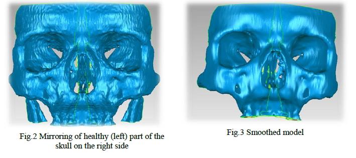
That’s where models of the skull and face are critical to planning surgeries.
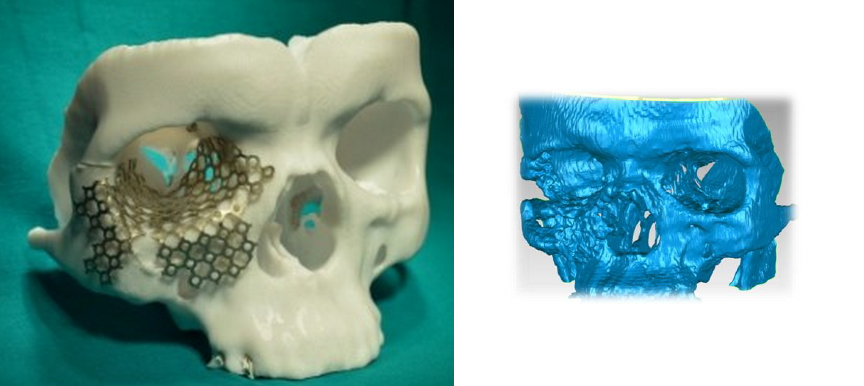
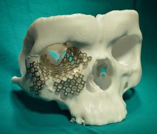
Researchers — from the Engineering Institute of the Kazan Federal University in Kazan, the Tatarstan Regional Clinical Cancer Center, the Russia Laboratory of Radiation Physics, and the Physical-Technical Institute of the Kazan Scientific Center in the Russian Academy of Sciences — built and printed a pair of 3D models of the front part of the skull to evaluate the effectiveness of its use in the planning of complex operations on cancer patients.
The team then built the models using a Stratasys Fortus 400mc and a 3D Systems ProJet 160 to evaluate the differences in materials the devices use. The ProJet 160 uses powdery VisiJet PXL, and the Fortus uses industrial-strength thermoplastics like ABS-M30.
Researchers say the models took approximately 14-15 hours to print and then spent 2-3 hours in a chamber containing a support removal solution. All in all, they say the functional anatomical models took less then 48 hours to complete and add that costs for planning were reduced by avoiding the pre-fitting of standard commercial products like grids and plates which are commonly used in planning surgeries.
Do you think 3D printing will become a critical tool for solutions to surgical problems and planning? You can weigh in with your thoughts on the ‘3D Printing Complex Facial and Skull Surgeries’ forum thread on 3DPB.com.