Researchers at the University of Bari have developed new methods of 3D printing for more effective assisted reproduction interventions and procedures for the protection of endangered species.
In vitro fertilization (IVF) is the most common assisted reproduction solution for couples facing fertilization problems. Since it was first applied successfully in 1978, more than 5 million babies have been born using IVF. Infertility impacts a significant portion of the population, with over 11% of the population or 6.7 million women worldwide unable to reproduce, as per the American Society of Reproductive Medicine.
In turn, methods that improve the viability and efficacy of IVF are of tremendous value. This is true not only for infertile couples, but also to researchers limited by conventional 2D methods, which have low reproducibility, and limited studies for validation in assessing or studying IVF. Such solutions also apply to domestic animals, the animal production industry, and in saving endangered species.
In their research, published in Plos One, an innovative bioengineering approach was used to encapsulate animal cells in a hydrogel microsphere. These microspheres were made using 3D bioprinting to obtain structures for culture in vitro, and are largely composed of water. An open source automated tool, Sphyga, was developed to generate spherical hydrogel spheres, using a syringe with the required shape, size and stability, and can be used to automate generation of alginate hydrogel microbeads.
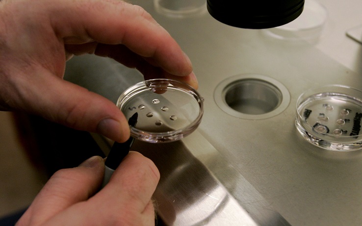
Embryos being studied for IVF. (Photo by Sandy Huffaker/Getty Images)
These microencapsulated eggs were found to have preserved structural and functional integrity, and have a higher development potential and viability as compared to eggs transferred conventionally, and the 3D bioprinting method—called 3D in vitro maturation (3DIVM)—was found to be highly reproducible and efficient. Researchers believe 3DIVM could become the preferred method in clinical and toxicological applications.

The Sphyga 3D bioprinting system developed for fabrication of hydrogel microbeads for use in IVF research (Image courtesy Plos One, Research at University of Bari)
At the Veterinary Medicine department at the University of Utrecht, researchers developed an oviduct-on-chip model to better study fertilization in humans and animals. Although they found certain 3D printing polymers are not viable for bioprinting and bioengineering due to toxicity, their results showed how important the 3D printed model was over traditional cell culture methods. The model was used to create conditions similar to that in the oviduct, considered more important than it was before in the fertilization process, using an air-liquid interface in a 3D printed system with filters for oviduct epithelial cells to grow.
This new method could also improve the effectiveness of IVF for animals such as horses and cows. In 2017, a paper published in Nature reported how scientists had successfully used 3D printing to make artificial ovaries, a gelatin-based bioprosthetic scaffold, to restore the fertility of infertile mice. Earlier in 2014, French researchers had patented a new way to study and examine in-vitro embryos, prior to implantation, ex-vitro – or via a 3D model and printed specimen that is based on a scan of the in-vitro embryos.