German researchers continue in the quest to improve processes in bioprinting and bone regeneration, sharing their recent study in ‘3D Printing of Piezoelectric Barium Titanate-Hydroxyapatite Scaffolds with Interconnected Porosity for Bone Tissue Engineering.’
Challenges seem to be synonymous with bone regeneration, one of the most serious obstacles for surgeons today attempting to treat patients requiring enhanced bone remodeling, repair, and growth. Tissue engineering is an extremely complex science, not only requiring enormous effort to keep cells alive and viable but also in finding suitable materials that offer the potential for repair strategies—and are biocompatible.
Other properties required for functioning bone grafts and compatibility include:
- Scaffold design
- Surface topology
- Chemistry
- Porosity
“In particular, the porosity of biomaterials plays an essential role in the context of osteointegration and osteoconduction and supports the migration of cells, capillary ingrowth and the transport of nutrients to cells. Functional bioinspired designs can be produced by utilizing advanced manufacturing techniques, such as electrospinning, freeze casting, sol-gel-techniques or additive manufacturing,” stated the authors.
“In particular, additive manufacturing, such as binder jetting, selective laser sintering or extrusion-based techniques became increasingly attractive based on their broad versatility and the ability to fabricate freely designed and patient-specific geometries.”
Conductive materials, and more specifically—materials like piezoelectric ceramics—are in a unique class of their own, and becoming increasingly popular in accompanying 3D printing technology due to their potential for transforming in shape, use with a variety of materials and techniques like bioprinting, and the ability to offer an electric response.
The authors point out that with additive manufacturing processes, they have the new capacity to make complex geometries with ‘enhanced osteogenic properties.’ 3D printed implants are customized, patient-specific, and can offer biocompatibility as well as stimulation due to the piezoelectric effect.
In this study, BaTiO3 powder was used for fabrication of scaffolds, with a particle size of d50 of <3 µm. Spray-dried hydroxyapatite powder with a grain size of d50 of ~40 µm was combined to create a BaTiO3/HA powder blend. Polyethylenmethacrylate was also added for greater stability in the scaffolding after 3D printing of the samples on a Voxeljet VX500.
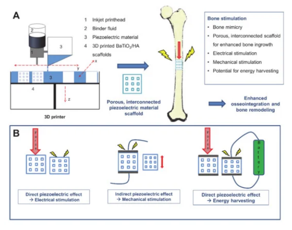
Overview of 3D printing of piezoelectric materials for bone stimulating implants. (A) shows exemplarily the binder jetting process used for the fabrication of piezoelectric scaffolds. (B) indicates different applications of the piezoelectric effect for bone stimulation. Piezoelectric implants have the potential to stimulate electrically (direct piezoelectric effect), mechanically (indirect piezoelectric effect) or could be used as an energy harvesting device to power other implants or sensors.

CAD Design and geometrical data of BaTiO3/HA composite scaffolds.
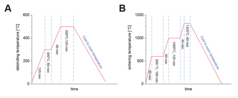
Applicated debinding, (A) and sintering curve, (B) for 3D printed BaTiO3/HA composite scaffolds.
The researchers then imbued the scaffolds with piezoelectric properties via a polarization formula comprised of electrodes connected to a high voltage power supply. Different settings were used:
“To find the best polarization parameters, the field strength, polarization time and polarization temperature were altered in 4 steps starting from 0.667 kV/mm to 1.25 kV/mm. The piezoelectric constant d33 of different polarized scaffolds (n = 5 samples for each group, full cylinder) was measured with the Berlincourt method using a d33 piezometer (PM300, PIEZOTEST, Singapore).”

SEM image of the BaTiO3/HA powder mixture used for the 3DP process (A, scale bar: 20 µm). Elementary classification by EDX spectroscopy for HA (B, scale bar: 90 µm) and BaTiO3 (C, Scale bar: 90 µm).
Many pores were present in the 100–200 µm range, offering favorable conditions for osteogenesis; however, the research team noted ‘very limited capability’ in terms of holding up under mechanical forces. The high porosity—and resulting brittleness—also made it difficult to attain necessary data.
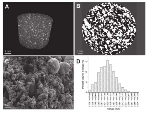
(A) Three-dimensional maximum intensity projection (MIP) of a BaTiO3/HA scaffolds with visible particles of different densities (scale bar: 2 mm); (B) The binarised cross-sectional microCT images reveal the number of pores (black) and provide the basis for a 3D calculation of porosity (scale bar: 1 mm). (C) The SEM images underline the results visible in the microCT of a highly porous network of particles which are roughly sintered. Large particles of HA are embedded in a percolating network of BaTiO3 particles through the whole scaffold (scale bar: 2 µm); (D) The pore size distribution of a 3D printed BaTiO3/HA scaffold.
“The compressive strength of 3D printed BaTiO3/HA scaffolds varied in a range of 50–370 kPa, resulting in an average compressive strength of 150 ± 120 kPa. Overall, the scaffolds were easy to manage and survived any transport and treatment,” concluded the researchers. “Nevertheless, a future aim for research is increasing the mechanical properties significantly by changing the sintering treatment or composition.
“The addition of further bioactive phases to the ceramic powder mixture will be investigated to tailor the bioactivity of the scaffolds and to potentially allow tailoring of the interface of BaTiO3/HA/X scaffolds to achieve increased mechanical performance. We show that the additive manufacturing of lead-free piezoelectric BaTiO3-based ceramics represents a promising approach to yield scaffolds of designed porosity, equipped with piezoelectric properties for enhanced bone regeneration.”
What do you think of this news? Let us know your thoughts! Join the discussion of this and other 3D printing topics at 3DPrintBoard.com
[Source / Images: ‘3D Printing of Piezoelectric Barium Titanate-Hydroxyapatite Scaffolds with Interconnected Porosity for Bone Tissue Engineering’]