In the recently published ‘Smartphone-based diagnosis of parasitic infections with colorimetric assays in centrifuge tubes,’ international researchers from Spain and the UK explore better ways to diagnose medical conditions in developing countries with few resources for such tools.
Although billions of consumers are using smartphones today, in nearly every recess of the world, most of us rarely take true stock of the power we are holding in the palms of our hands. These devices that carry so much of our personal information and may only be used for sending a few texts or making a few calls each day are actually powerful microprocessors capable of performing a vast number of complex—and extremely helpful—tasks, as well as sharing enormous amounts of data.
Here, the authors put the smartphone to the test for diagnosing parasitic infections, opening our eyes to what a critical role the camera can play in bioanalytical and diagnosis applications. Focusing on the diagnosis of Leishmaniasis and Chagas disease, the team explains that these illnesses can be severe—and so much so that they are responsible for making millions of people sick. With the development of an affordable, easy-to-use colorimetric assay for diagnostics of Leishmaniasis and Chagas disease, the researchers hope to make an impact of more remote areas of numerous countries.
“In this study, the proposed smartphone-based diagnostic platform has been shown to be capable of making analysis of millimetric colorimetric arrays, as those fitting into the Spin-Tube. Although in the case of the Spin-Tube platform published by our group recently, where the spot pattern recognition was undertaken by naked-eye (due the simplicity of the colorimetric pattern to needing to be analyzed), further development of an automatized image capture system, such as a smartphone is an attractive option,” state the researchers.
This innovative new device offers the following advantages for patients, technicians, and doctors:
- Reduction in human error as many staff may be untrained
- Creation of a permanent record for doctors to review as necessary
- Enabling of quantitative assessments through spot color intensity measurement
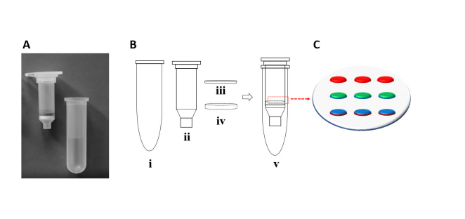
A) Spin-Tube device prototype. B) Plastic components for the Spin-Tube fabrication. C) Amide bond formation between pre-activated carboxylic acid groups of nylon membranes and primary amine groups of
abasic probes (PNA1 and PNA2). Graphic layout of the array: In red, 3 biotin-labelled DNA oligomer controls; in green, 3 spots of abasic PNA2; in blue, 3 spots of abasic PNA1.
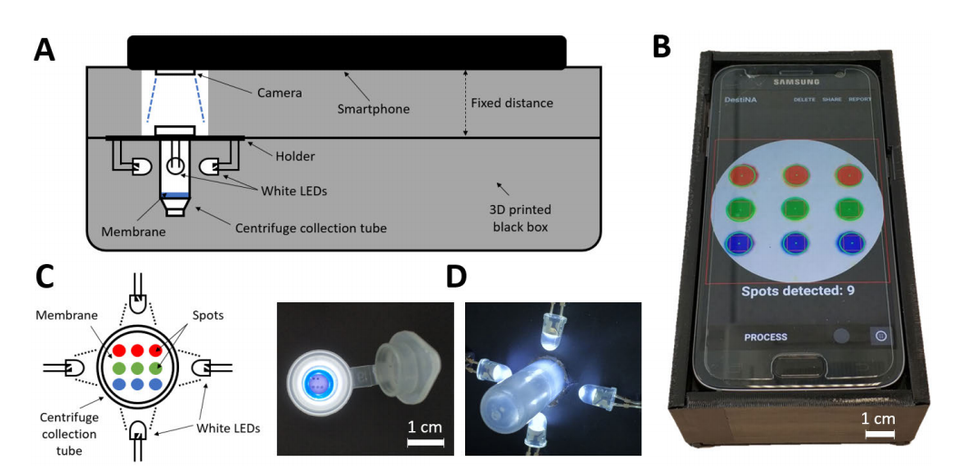
A) Lateral schematic view and B) photograph of the complete setup composed of the 3D printed accessory, the illuminated centrifuge collection tube and the smartphone. C) Top schematic view and D) photographs of the centrifuge collection tube with the white LEDs.
The authors also 3D printed an affordable accessory made of plastic for adding illumination. A software program was also programmed for the smartphone, sensing color-coordinates of circular-shaped spots in each array pattern.
“The app consists of several screens that guide the user through the steps required for the acquisition and processing of a test result image,” explained the researchers.
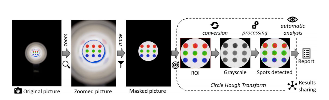
. Image acquisition and processing workflow in the smartphone application.
Numerous tests were performed to evaluate system performance, verify the application overall, including testing its limits.
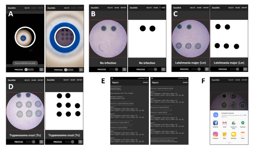
A) App screenshots where the user can fit the template grid (white circle) with the centrifuge collection tube after taking a photo. Examples of results of diagnostic recognition (detailed and simplified views) upon the completion of the processing for the three possible cases: B) Absence of infection; C) pathogen Leishmania major; and D) pathogen Trypanosma cruzi. E) Example of the report provided by the app containing information about the recognized spot array. The report is presented in a plain text viewer integrated in the app. F) Both the report and the processed image can be shared through social networks, email and various messaging services through the ‘‘Share’’ button in the top menu of the application. In this study, the spotting was carried out with 2 controls biotin-labelled DNA oligomer spots rather than the 3 of Fig. 1C.
“This system has been tested for detection of parasitic diseases of the family Trypanosomiases, which are responsible for devastating diseases in humans, dogs as well as livestock. In this context, the smartphone-based platform that has been developed analyses the image and converts the panel of spots into a Trypanosomatid species,” concluded the researchers. “Given the importance of such assays in developing countries, the mobile phone imager application has been designed to be especially user-friendly for its use by untrained personnel. Recorded results can be immediately transmitted to reference clinicians from remote locations for advice and treatment decisions.”
Smartphone devices have been developed for a wide range of applications so far, many of which are helpful to medical professionals and patients in developing areas, from testing for malaria to high blood pressure, and even diagnostics for pancreatic cancer.
What do you think of this news? Let us know your thoughts! Join the discussion of this and other 3D printing topics at 3DPrintBoard.com.
[Source / Images: ‘Smartphone-based diagnosis of parasitic infections with colorimetric assays in centrifuge tubes’]