
Medical simulation can be invaluable for residents in training, although before the advent of availability with 3D printing, there was not quite as much accessibility—or possibility; in fact, cadavers are still considered the ‘gold standard’ for training with simulators, but they are more expensive, high-maintenance, and highly regulated.
Looking at the trend toward creating simulators that can mimic the feeling of penetrating cortical bone into cancellous bone during spinal instrumentation, the authors created three lumbar vertebrae and 20 C2 vertebrae models, using an Ultimaker S5 3D printer with PLA filament, and PVA (polyvinyl alcohol)—used to imitate cancellous bone inside the shell. While PVA is a plastic that tends to be spongy and water-soluble, it is generally not used in creating stand-alone structures, and has had limited use. With the accompaniment of PLA, however, parts are less brittle and exhibit greater ductility. The researchers state that here, the two materials are suitable for creating simulated bone.

(A) Solid view and (B) layered view demonstrating the planned infill matrix of the PVA (yellow, black
arrow) and the outer cortical shell (red surface)
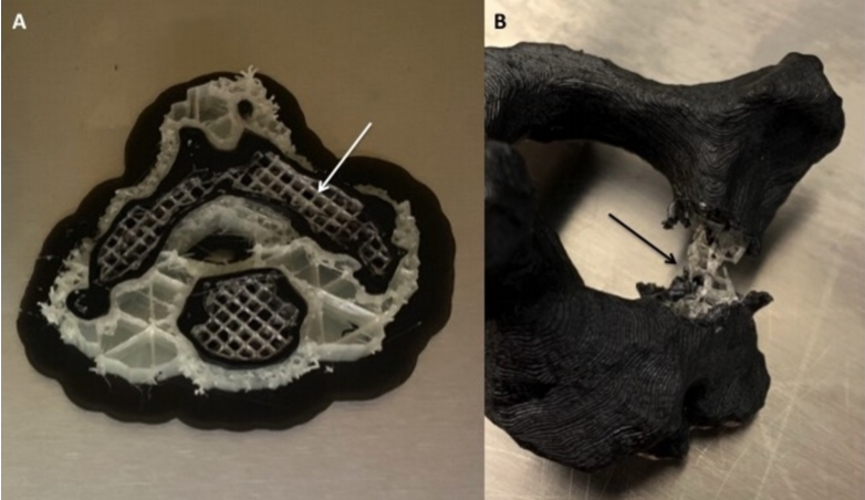
(A) The interface of the PLA (black) and PVA (white matrix, white arrow) is shown during a printing session. (B) The outer shell of “cortex” (PLA) is removed from the pedicle of a finished model demonstrating the inner matrix of PVA (black arrow). An advantage of dual-extrusion 3D printers is the ability to change material deposition within the same print.
The researchers reported good success with all the models. There were no failures reported in either hardware or materials, and the PLA/PVA models proved to be superior over the single material models, and in tactile feedback, the researchers said that they ‘accurately represented the sensation of in vivo instrumentation during pedicle probing, pedicle tapping, and screw placement.’
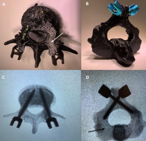
(A) L3 vertebrae with PLA shell removed demonstrating the spongy PVA matrix (white arrow). Pedicle screws are seen within the matrix. (B) Laminar screws placed in the C2 experimental model without model failure. (C & D) Fluoroscopic images obtained post instrumentation. The PVA infill matrix can be seen under X-ray (black arrow).
Previously, popular 3D printed materials for fabricating vertebral bony anatomy for biomechanical testing and instrumentation have been used with a wide range of different results:
- A variety of resins
- Acryl butadiene styrene (ABS)
- Nylon
- Polylactic acid (PLA)
- Thermoplastic urethane (TPU)
“Another limitation of most previously published models is their constraint of a one material print. Literature suggests that decreasing the print infill percentage adequately simulates the density change of cortical bone to cancellous bone. These findings have been quantified with regards to subjective user feedback and biomechanical stress with good results,” stated the researchers. “A possible limitation of this modeling technique is the ultrastructure of the print does not completely simulate the complex characteristics of human vertebrae.
“Bone strength is composed of two properties: bone mass, or quantity of material, as well as the bone quality, or mechanical properties of the material. Previous spinal models have incorporated these parameters individually, but not collectively.”
Shells printed with dual materials were selected because of anatomical differences and instrumentation placement methods and were limited to only the lumbar and cervical forms. Ultimately, the PLA/PVA method was promising for use in both educational and biomechanical simulators.
“This has large implications for the spinal instrumentation industry as well as resident training. This concept and design of simultaneous multi-material printing using our unique infill algorithm have not been yet reported in the medical literature,” concluded the researchers. “Further educational and biomechanical testing on our design is currently underway to establish this printing method as a new standard for spinal biomimetic modeling.”
Medical models overall have become extremely helpful in medicine, and while simulators may sometimes be larger and more complex in design, smaller models are often used for diagnosing conditions like tumors, training and surgical planning with 3D printed models of the brain, and even those that allow parents to plan for babies with medical issues. Find out more about how 3D printing is making a positive impact with progressive simulators here. What do you think of this news? Let us know your thoughts! Join the discussion of this and other 3D printing topics at 3DPrintBoard.com.
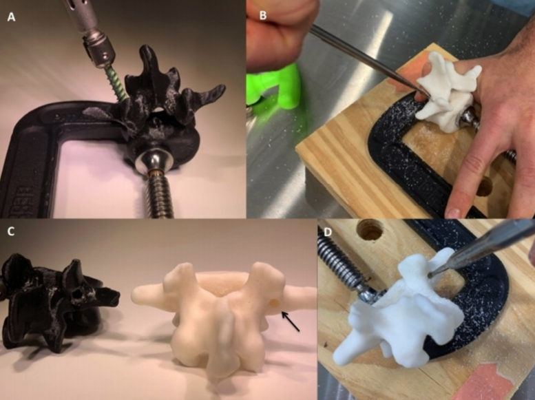
(A) The L3 PLA/PVA experimental model was successfully probed, tapped, and instrumented with a pedicle screw. (B) The resin print pedicle was unable to be accessed manually with the curved probe due to its solid properties. (C) The resin print (white) is shown to be completely solid (black arrow) when a pilot hole is drilled (D) with a high-speed burr. The pedicle of the resin model cracked after it was drilled and tapped.