![]()
The use of scaffolds has been rife with challenges, and especially in guided bone regeneration (GBR). Problems include issues with handling in clinical environments, erratic degradation, lack of stability in cell growth, and more. The researchers in this case embarked on a study of scaffolds created through melt electrospinning writing (MEW) and consequent 3D printing, creating complex scaffold geometrics that can be fabricated specifically to bone tissue requirements—usually in maxillofacial applications.
“In general, membranes that are currently used for maxillofacial applications, such as GBR, can be broadly divided into resorbable and non-resorbable categories,” state the researchers. “Membranes of the latter category offer good biocompatibility and high mechanical stability. Thus, they suit very well as placeholders and barriers in GBR. On the other hand, non-resorbable membranes require a second operation for their removal, pose a risk of mucosal perforations due to their high level of stiffness and therefor go along with higher morbidity, increased costs and increased expenditure of time.”
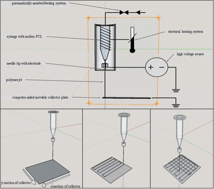
Design of MEW device and schematic of the MEW process of the scaffolds
Electrospinning offers a comparatively easy way to make scaffolds necessary to biomedical applications, employing a charged polymer jet pushed out from a spinneret and ‘drawn’ toward an area well fibers begin to form into a structure. For creating polymers, both solution electrospinning and MEW are used.
MEW allows the operator to place fibers exactly, and without chemicals and solvents, meaning there is also no potential for solvents to be left in the scaffold.
“Process parameters allow for a controlled fiber placement, which makes the computer-aided design- and manufacturing (CAD/CAM) of 3D scaffolds feasible. Cell growth on scaffolds can thus be optimized by varying pore sizes and interconnectivity,” explain the researchers.
All scaffolds for the study were made of medical-grade PCL and the researchers were able to use a MEW device that was customized by the Department for Functional Materials in Medicine and Dentistry Würzburg University Hospital. Cells were assessed and quantified, and then washed, sealed, and neutralized. Testing revealed good viability and proliferation.
“The architecture of scaffolds, size, morphology and interconnectivity play an important role in the sufficient regeneration of bone,” state the researchers.
“Such scaffolds/membranes would prove beneficial, especially in the field of oral and maxillofacial surgery, where impaired wound healing in the alveolus region oftentimes poses problems. Furthermore, a pre-implantological use of these membranes seems suitable. In atrophic regions of the alveolus in which elaborate bone augmentation is necessary, coverage with membranes that additionally promote bone healing should prove a promising approach to improved GBR.”
End results showed not only good viability but also protein concentration, and cell number.
“These well-defined 3D scaffolds consisting of medical-grade materials optimized for cell attachment and cell growth hold the key to a promising new approach in GBR in oral and maxillofacial surgery,” concluded the researchers.
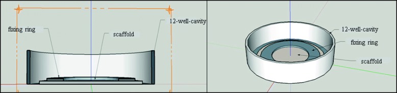
Schematic of cell culture experiments on the scaffolds
3D printing has lent so much to the world of innovation, from aerospace to fashion and beyond, but this technology has had significant impacts in medicine, and in areas that are vital but that many of us rarely think about or even know about, such as scaffolding.
What do you think of this news? Let us know your thoughts! Join the discussion of this and other 3D printing topics at 3DPrintBoard.com.
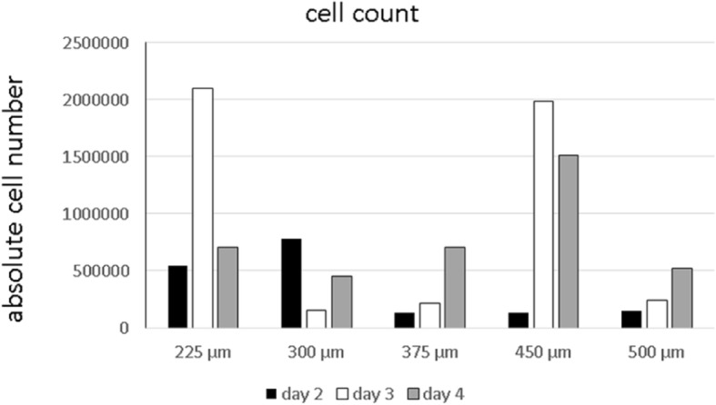
Cell count of MG63 cells seeded on MEW box structured PCL scaffolds of different box-sizes after 2, 3 and 4 days of cell culture