Hydroxyapatite is a naturally occurring mineral that is the main inorganic constituent of both tooth enamel and bone. In a paper entitled “Optimization of extrusion based ceramic 3D printing process for complex bony designs,” a team of researchers describe how they 3D printed the material, studying the effects of different parameters, and finally 3D printed a patient specific bone graft.
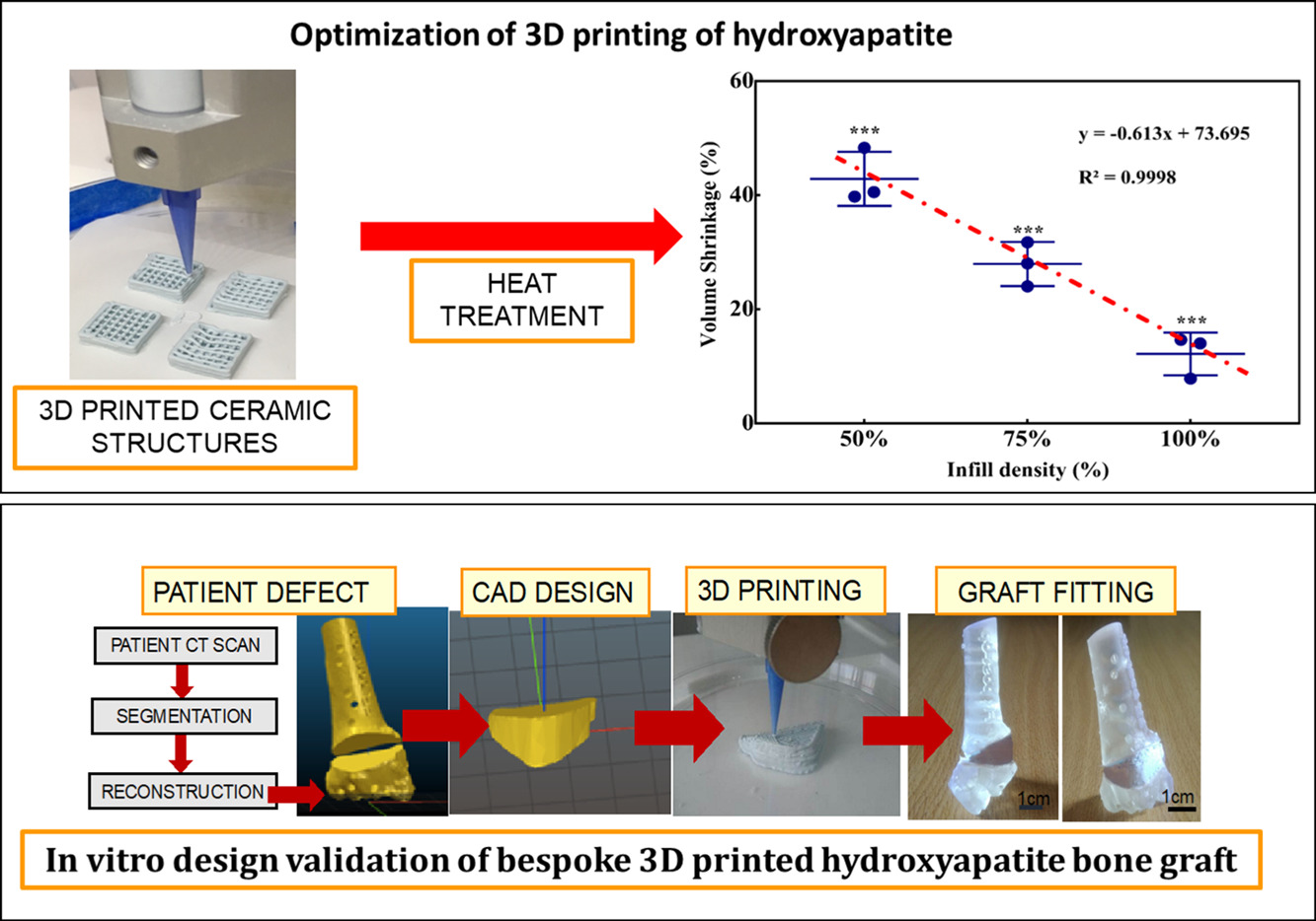
The researchers began by 3D printing several cube-shaped scaffolds using INKREDIBLE+ 3D printing ink from CELLINK. After printing, the samples were dried, sintered and cooled. Dimensions of the scaffolds were taken using calipers, and mechanical testing was carried out to test the compressive strength of the scaffolds. The samples were then analyzed using scanning electron microscopy, and micro CT scanning was carried out to take a non-destructive qualitative image of the scaffolds and to carry out a quantitative 3D analysis of the porosity, volume fraction and degree of anisotropy.
A case study was then performed.
“Briefly, CT scan of distal radius malunion was reconstructed and the required clinical defect and graft were reconstructed and designed using CT imaging…from the elbow to the carpometacarpal joints was performed,” the researchers state. “The DICOM files were imported into medical image processing software…to segment the CT images and generate 3-dimensional virtual models of both forearm bones. Consequently, a virtual osteotomy was performed to produce a precise correction of angular and rotational malalignment of the radius.”
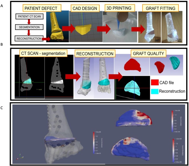
The 3D model of the patient defect was 3D printed with clear resin using a Formlabs Form 2 3D printer. The CAD of the defect graft was scaled up to compensate for post-sintering shrinkage, and was then 3D printed using hydroxyapatite ink. After sintering, the graft was glued inside the defect and a CT scan was taken of the entire construct.
The researchers discovered during the process that post-sintering shrinkage is proportional to infill density.
“The average external volume after sintering is less for 50% and 75% infill when compared to 100% infill density,” they explain. “…This relationship between volume shrinkage and infill density can be used to accurately predict level of shrinkage for 3D printed geometries using HA ink developed in this study. This thus allows us to use this information to scale up geometries during design process, so that accuracy of final print, post-sintering process is maintained.”
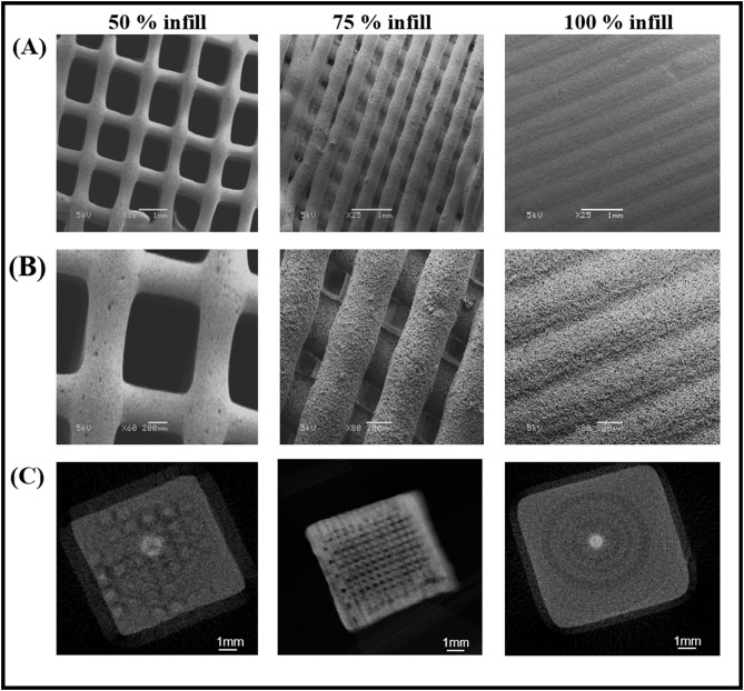
Various parameters, including print speed, extrusion pressure, and ink viscosity, all contribute to the porosity of the scaffolds before sintering. After the binder is eliminated from the scaffold matrix, the rise in temperature causes particles to vibrate and come closer to each other, reducing porosity and increasing stability.
“Understanding relationship between shrinkage of 3D geometry post sintering process of HA based 3D printed structure provides a predictable correlation between design files and final processed objects so that patient specific bone defects can be printed from pre-operative CT scans,” the researchers conclude. “This study has implications for materials and 3D design process optimisation for clinical applications. Patient specific bone graft production has potential to reduce need for harvesting autograft and decrease surgery time, whereby impacting overall outcome of the surgery and related costs.”
Authors of the paper include Roopavath Uday Kiran, Sara Malferrari, Annemieke Van Haver, Frederik Verstreken, Subha Naryan Rath and Deepak N. Kalaskar.
Discuss this and other 3D printing topics at 3DPrintBoard.com or share your thoughts below.