In theory, a team of researchers led by John R. Shaffer, Ekaterina Orlova, and Myoung Keun Lee from the University of Pittsburgh have released a set of convincing evidence that could be used to justify that genes influence the formation and development of facial appearance.
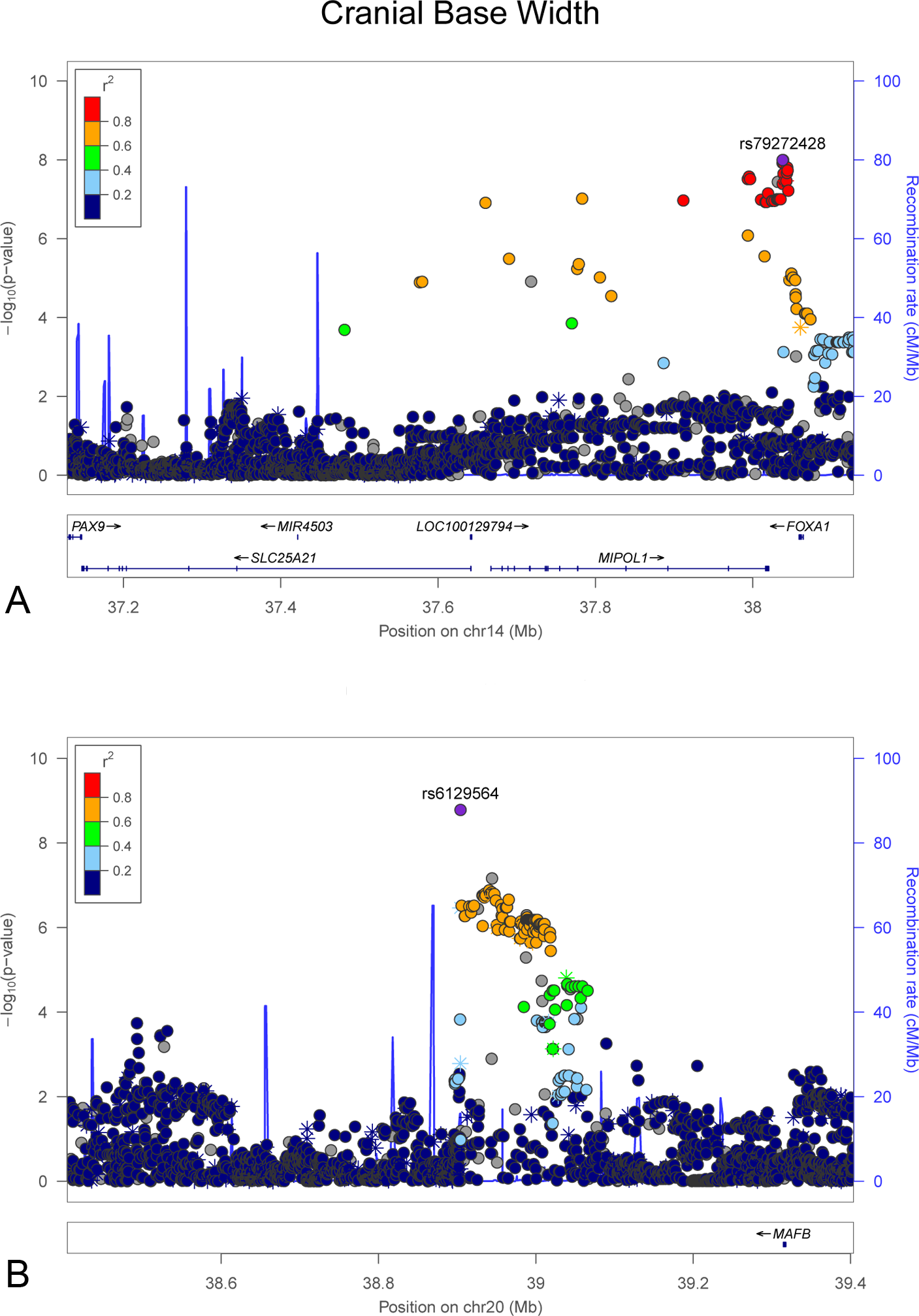
With 3D imaging, researchers at the University of Pennsylvania gathered the 3D surface images of 3,118 individuals of European ancestry. All scans were analyzed by the researchers, who observed genome-wide associations in the facial features. Researchers discovered that associations existed between genes and the formation of cranial base (skull structure), intercanthal width (corner of the eye; where the eyelids meet), and nasal width.
Various data sets from the observational study led to a conclusion that certain genes play crucial roles in the development of craniofacial features, significantly affecting facial structures and features. In addition to the 3D imaging-based observational study, researchers also performed a test on genotype-phenotype associations, which further suggested that harboring genes impact variation in facial features.
The University of Pennsylvania’s team of researchers emphasized that the observations and discoveries from the 3D surface images will help the understanding of facial morphology, thus revolutionizing the field of abnormal facial morphogenesis.

3D Imaging Technique; Canfield Scientific
“Improved understanding of the genes associated with facial morphology in healthy individuals can provide insights into the pathways and mechanisms controlling normal and abnormal facial morphogenesis,” read a section of the study.
“Our ability to connect specific genetic variants to ubiquitous facial traits can inform our understanding of normal and abnormal craniofacial development, provide potential predictive models of evolutionary changes in human facial features, and improve our ability to create forensic facial reconstructions from DNA,” the authors stated.
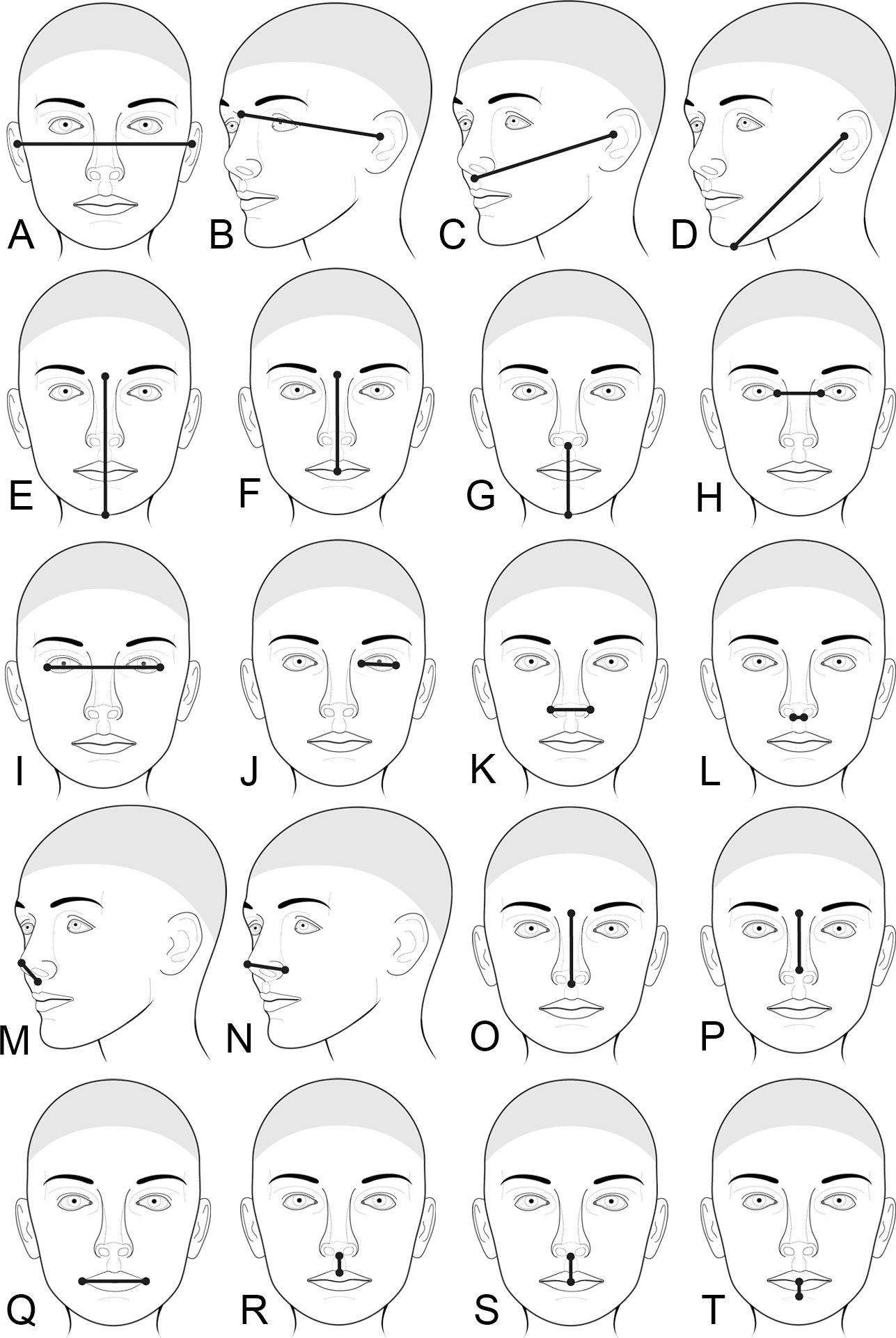
As seen in the study infographic at right, each unique 3D image of the facial structures of individuals of European descent allowed the researchers to produce a comparison chart between evolutionary changes in facial features, which makes it simpler for researchers in the field to understand the relationship between genetic variance and facial structure.
In addition to Shaffer, Orlova, and Lee, contributing authors in full included Elizabeth J. Leslie, Zachary D. Raffensperger, Carrie L. Heike, Michael L. Cunningham, Jacqueline T. Hecht, Chung How Kau, Nichole L. Nidey, Lina M. Moreno, George L. Wehby, Jeffrey C. Murray, Cecelia A. Laurie, Cathy C. Laurie, Joanne Cole, Tracey Ferrara, Stephanie Santorico, Ophir Klein, Washington Mio, Eleanor Feingold, Benedikt Hallgrimsson, Richard A. Spritz, Mary L. Marazita, and Seth M. Weinberg. Discuss this in the Genetics Study forum at 3DPB.com.

3D Imaging Technique; Canfield Scientific