
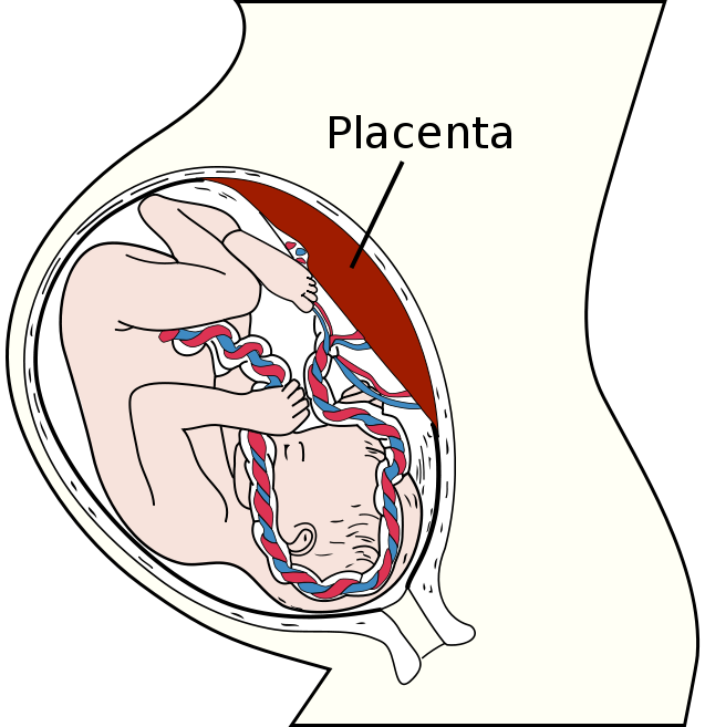
There are some medical theories that suggest that one of the causes of preeclampsia lies with a type of special cell in the placenta called trophoblasts. In a healthy placenta, the trophoblasts attach themselves to the uterine wall and then start to grow into the tissues of the uterus during the first stage of pregnancy, essentially bonding both organs together. At that point they connect to the mother’s blood vessels, which plays a vital role in helping to nourish and encourage growth in the fetus. The theory is that if the trophoblasts do not migrate normally it could lead to poor placental perfusion, the insufficient blood flow between a placenta and a uterus that causes preeclampsia.
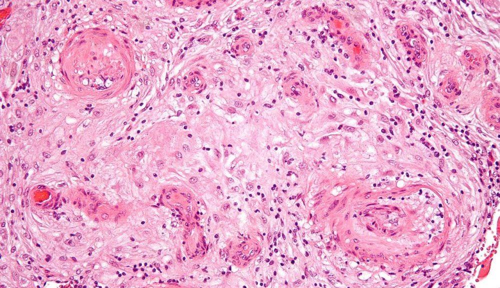
Cells in the placenta during preeclampsia are inflamed due to hypertension.
In a new study that was published in American Chemical Society (ACS) Biomaterials Science & Engineering, the scientists detail their research which involved using the bioprinted model to observe the migration of trophoblasts cells form the placenta to the uterus. The bioprinted placenta model created by the research team contained key cellular, biochemical, and extracellular matrix components that accurately simulated the process of trophoblast migration. This is the first time scientists were able to successfully 3D bioprint a placenta and test cell migration of this type, and it could lead to several life-saving medical treatments.
“Our study provides a proof of concept that a 3D bioprinted placenta model is a viable way to study and understand the dynamics of cell migration in the formation of the placenta. What we have learned from this initial study is a significant step toward understanding the cause of preeclampsia and a potential therapy,” explained the chair of the Fischell Department of Bioengineering and the University of Maryland, John P. Fisher.

The bioprinted placenta model allowed trophoblasts to be monitored during migration.
The team was able to successfully use the 3D bioprinted placenta model to track the results of introducing epidermal growth factor (EGF), a peptide that stimulates cell growth, proliferation, and differentiation, on trophoblast migration. The results strongly suggest that introducing the EGF to the placenta had a positive effect on the migration of the trophoblasts. While there is still more testing required, it seems that EGF may be a potential therapeutic treatment for preeclampsia. The reason that the 3D bioprinted model was able to provide such valuable data is the fact that the scientists were able to learn far more about the dynamic behavior of the trophoblast cell movements than more traditional 2D models.
When a 2D model is used researchers can only tell that a cell has moved, but with the 3D model they were able to see how it moved, where it moved and if a group of cells moved together. According to their research paper, the 3D bioprinted placenta model is a promising first step to developing a more sophisticated placenta model that could be bioengineered as a powerfultesting and research tool. Being able to accurately simulate the biological workings of an organ like a placenta could help them develop new treatments for preeclampsia and other similar placenta-related conditions including placenta accreta and placenta previa.
The study, ‘Development of a 3D Printed, Bioengineered Placenta Model to Evaluate the Role of Trophoblast Migration in Preeclampsia‘, was authored by Che-Ying Kuo, Avinash Eranki, Jesse K. Placone, Kelly R. Rhodes, Helim Aranda-Espinoza, Rohan Fernandes, John P. Fisher, and Peter C. W. Kim.
Do you know anyone who suffered from preeclampsia who would find this information interesting and helpful? Let’s discuss over in the 3D Printed Bioengineered Placenta Model forum thread at 3DPB.com.