Industrial 3D printing has been around since the 1980s and was initially known as rapid prototyping because of its most popular application – making prototypes for manufacturing. The term additive manufacturing includes such technologies as stereolithography, fused deposition modeling (FDM), laser sintering, and electron beam sintering. Recent advances in additive manufacturing reveal that bioprinting, i.e., the additive manufacturing of tissues and organs, is about to open up a whole new realm of possibilities. Though this technology has much in common with traditional additive manufacturing, and indeed adopts certain elements, there are a number of unique technical and legal challenges to implementing the use of bioprinted materials.
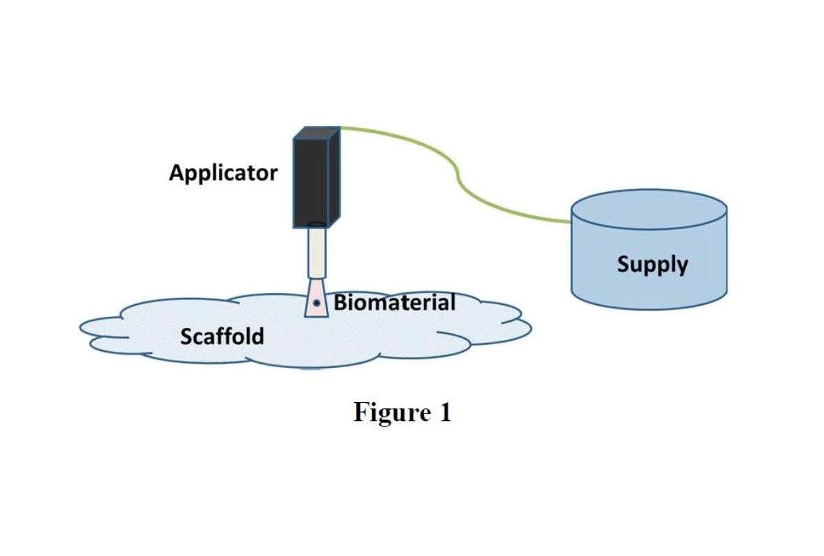
First, let us talk some basics. As shown in Figure 1, in order for additive manufacturing of biomaterials to work, the supply, applicator, and support structure must be constructed so that the biomaterial remains viable before, during, and after the construction of the tissue and/or organ. The biomaterial must also be able to thrive and grow in the environment it is intended for (after application). The high temperatures associated with traditional FDM, for example, could never work because the biomaterial would be destroyed. Further, something must hold the biomaterial together to shape it for its application, much like a support structure in traditional additive manufacturing.
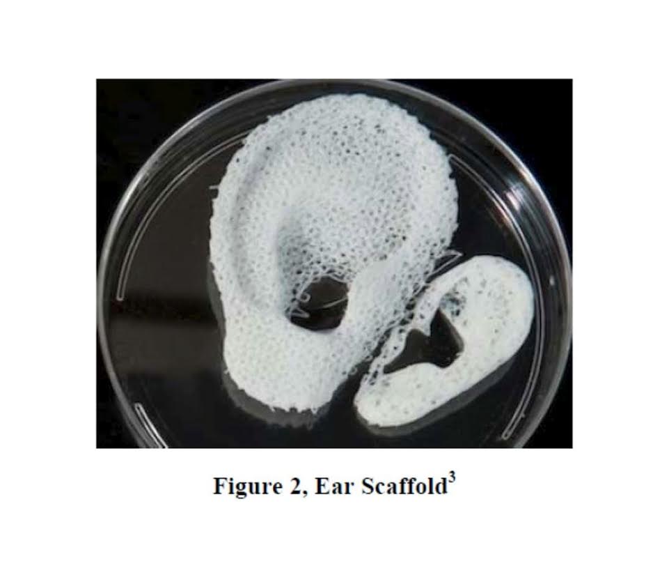
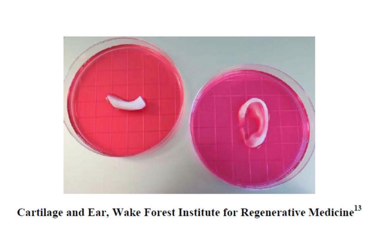
The other challenge is creating equipment that can deliver the biomaterial onto or into the scaffold. Wake Forest Institute for Regenerative Medicine at Wake Forest Baptist Medical Center has been a leader in the research in this field. Wake Forest has received funding from the Armed Forces Institute of Regenerative Medicine, a federally funded effort to apply regenerative medicine to battlefield injuries. The researchers have developed a custom-designed 3D printer and have printed ear, bone, and muscle structures.(9,10) These structures have been implanted in animals, matured into functional tissue and even developed a system of blood vessels. The printer can fabricate stable, human-scale tissue of any shape. The correct shape is achieved by converting clinical imaging data of an anatomical defect into a computer model to program control of the motions of the printer nozzles, which dispense the cells to discrete locations. With further development, “this technology could potentially be used to print living tissue and organ structures for surgical implantation.”(11)
The system, known as the “Integrated Tissue and Organ Printing System,” was developed over a 10-year period. The system deposits both biodegradable, plastic-like materials to form the tissue “shape” and water based gels that contain cells. A major challenge of tissue structures is to ensure that the implemented structures live long enough to integrate with the body. This was addressed by creating a hydrogel that holds the cells and a lattice structure of micro channels that allows nutrients and oxygen from the body to provide nutrients until the tissue regenerates its own system of blood vessels.(12)
The actual use of such 3D printed biomaterials on human beings is not that far away, though the regulatory framework of the United States Food and Drug Administration (FDA) presents additional challenges for tissues and organs as opposed to surgical implants which are made of existing approved and clinically accepted materials (such a titanium, steel, certain plastics, etc.). A party seeking to obtain regulatory approval for a device constructed from existing approved materials can typically streamline the approval process through the Premarket Notification Procedures under 510K. Biomaterials and/or organs, however, will need to proceed through the full approval process, meaning that there will have to be animal studies, clinical trials, an IRB (Independent Review Board), and proven results, prior to market approval, all of which is very expensive.
Currently, the FDA is working with the AM industry to develop new tools, standards, and approaches for the FDA to assess the safety, quality, efficacy, and performance of FDA-regulated 3D printed products. In its Department of Health and Human Services Justification of Estimates for Appropriations Committees for the 2015 Fiscal Year, the FDA stated that it is currently identifying medical device 3D printing standardized terminology, regulatory concerns, and developing quality control tests. The FDA further announced that it has identified how 3D printing techniques and processes affect the strength and durability of materials used in medical devices.
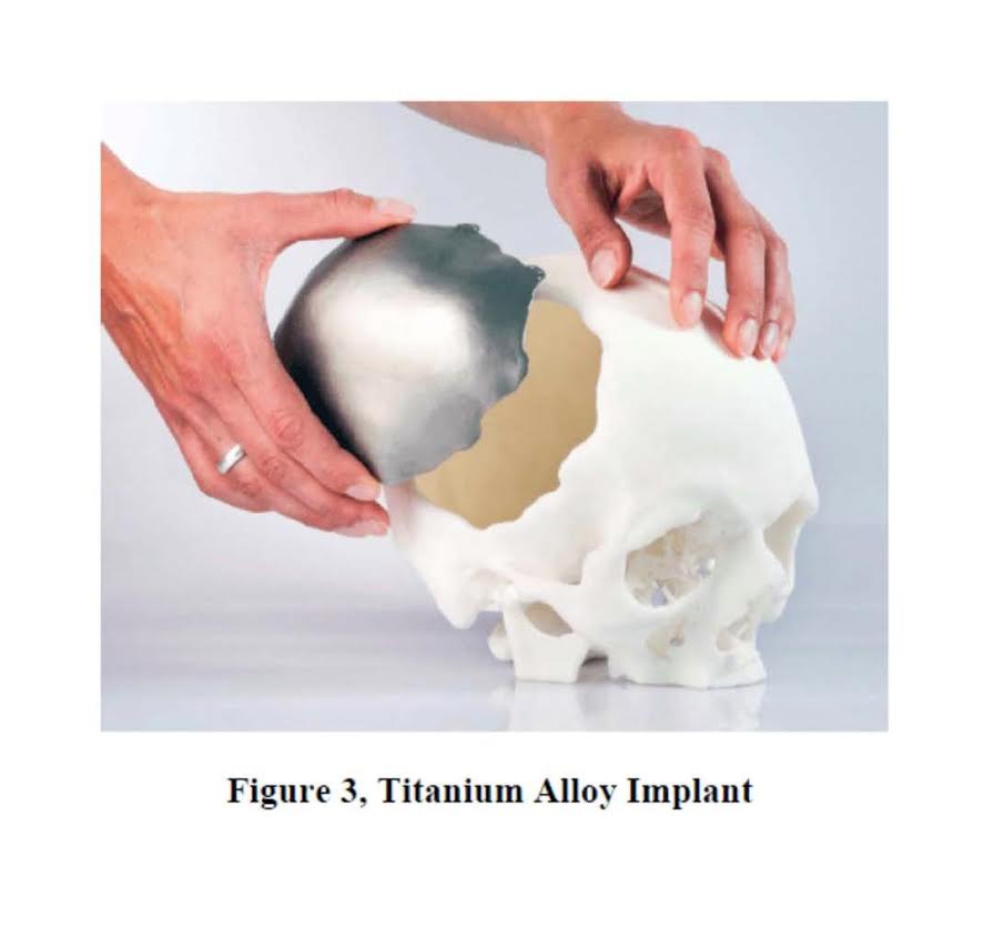
Europe has also approved the use of 3D printed materials as an implant. CEIT Biomedical Engineering, a Slovakia-based company, obtained EU approval for an implant constructed from a titanium alloy fabricated on an EOS laser metal sintered machine (see below, Figure 3). The use of known materials, such as the titanium alloy, presents less of a challenge for FDA approval.
William J. Cass, Esq. and Sandra L. Shaner, Ph.D.
William J. Cass is the Co-Chair of the Additive Manufacturing Practice Group of Cantor Colburn LLP located at 20 Church Street, 22nd Floor, Hartford, CT 06103-3207. Dr. Sandra Shaner holds her Ph.D. in physical chemistry and practices in the Chemical, Materials, and Life Sciences group within the firm.
(2) – Murphy SV & Atala A, 3D Bioprinting of Tissues and Organs, Nature Biotechnology, 2014, 32(8):773-785.
(3) – Downloaded from Wake Forest Institute for Regenerative Medicine Web Site https://www.wakehealth.edu/WFIRM/
(4) – Murphy SV & Atala A, 3D Bioprinting of Tissues and Organs, Nature Biotechnology, 2014, 32(8):773-785
(5) – Park SY, et al. Tissue-Engineered Artificial Oesophagus Patch Using Three-Dimensionally Printed Polycaprolactone with Mesenchymal Stem Cells: A Preliminary Report. Interact CardioVasc Thorac Surg 2016; doi:10.1093/icvts/ivw048.
(6) – Liu, A., et al. 3D Printing Surgical Implants at the Clinic: A Experimental Study on Anterior Cruciate Ligament Reconstruction. Sci. Rep. 6, 21704; doi: 10.1038/srep21704 (2016).
(7) – Jung, JW, et al. Computer-Aided Multiple-Head 3D Printing System for Printing of Heterogeneous Organ/Tissue Constructs. Sci. Rep. 6, 21685; doi: 10.1038/srep21685 (2016).
(8) – Peloso A, et al. Stem Cell Research & Therapy (2015) 6:107 DOI 10.1186/s13287-y.
(9) – “Scientists Prove Feasibility of “Printing” Replacement Tissue”, Wake Forest Baptist Medical Center, News Release February 15, 2016. https://www.wakehealth.edu/News-Releases/2016/Scientists_Prove_Feasibility_of_“Printing”_Replacement_Tissue.htm
(10) – Kang H-W, et al., A 3D Bioprinting System to Produce Human-Scale Tissue Constructs with Structural Integrity, Nature Biotechnology 34, 312–319 (2016).
(11) – “Scientists Prove Feasibility of “Printing” Replacement Tissue”, Wake Forest Baptist Medical Center, News Release February 15, 2016.
(12) – Id.
(13) – https://www.wakehealth.edu/WFIRM/ [last accessed 4/28/16].
(14) – Jung, JW, et al. Computer-Aided Multiple-Head 3D Printing System for Printing of Heterogeneous Organ/Tissue Constructs. Sci. Rep. 6, 21685; doi: 10.1038/srep21685 (2016).
(15) – Qu M, et al. Influence of Cell Printing on Biological Characters of Chondrocytes, Int J Clin Exp Med 2015;8(10):17471-17479.