
With 3D printing, the researchers have been able to fabricate the following:
- Femur
- Tibia
- Patella
- Knee reconstruction guide

All of this and more has been outlined in the paper, ‘A Novel Approach for Patellofemoral Tracking Using a Knee Model Reconstructed with a Three-Dimensional Printer,’ by Gian Luca Gervasi, Roberto Tiribuzi, Anastasios Georgoulis, Cerulli Giuliano, and Marco Freddolini. The research was recently published in 3D Printing and Additive Manufacturing.
With this model, the researchers employ all the benefits of 3D printing from customization and self-sustainability in research, to speed in production, and affordability. In their paper, they detail how they were able to use MRI scans from a patient’s knee to 3D print models of the three main parts of the knee, along with the guide which assisted in putting them together with artificial ligaments and a tendon. With these artificial ‘knee parts’ they were able to see what would happen—and how—as they applied force at varying degrees.
“The quadriceps tendon was simulated using a polyvinylchloride cord attached to the tibial insertion and the patella. The model was fixed to a tensile test machine and four static tests were performed by applying 200 N load in the proximal–distal direction through the cord at 30°, 60°, and 90° of knee flexion,” state the researchers in their paper. “The position of the patella was measured using a motion-tracking system with a custom-made navigation system.”
This research and the usefulness of the models is important because ‘patellar maltracking’ is the cause of knee pain, abnormal movement, and numerous joint disorders that lead to pain and mobility issues.
“…it is important to evaluate the kinematics of the patellofemoral joint in subjects with patellar instability. It is challenging to use in vivo kinematic analysis methods normally used for a precise analysis of lower limb joint motion because the data acquired with the motion-tracking system (e.g., optoelectronic system) are influenced by the relative movement of the markers on the skin that introduces errors in the recorded data,” state the researchers.
They exported the file data from the MRI into a 3D model with the use of FreeCAD 22 and then 3D printed on a MakerGear 3D printer. Once the model was complete, the researchers evaluated it with a compressive loading test. The researchers stated that three identical patellae were reconstructed with the same parameters used for the model patella, and then a compressive loading test was performed with an Instron 5965 materials-testing machine (Instron, Inc.).
“Neoligaments artificial ligaments (Xiros) were used as anterior cruciate ligament, posterior cruciate ligament, medial collateral ligament, and lateral collateral ligament to mimic the stability of the real knee. For this preliminary study, we chose only these principal ligaments that are sufficient to guarantee the stability of the knee,” state the researchers. “The ligaments were fixed to their insertion points, following anatomical landmarks at their isometric positions and using MRI to better identify insertion points.”
They found in evaluating the models further that the position of the patella for the preliminary study indeed followed patterns similar to what was found in ‘similar static and dynamic studies’ when employing cadavers for research. With patellar tilting being another important parameter to evaluate knee stability and different conditions, they evaluated its position. The patella tilted progressively in the lateral direction and showed a pattern similar to those observed for static and dynamic studies of cadaver’s knees. Patellar spin and flexion data were also consistent with in vitro studies.
The results of the study overall were very positive in that the 3D printed model, fabricated at a beneficially low cost was able to simulate patellar behavior in a more than sufficient manner. With such success, it was decided that researchers would be able to use the models to make customized surgical treatment plans for patients.
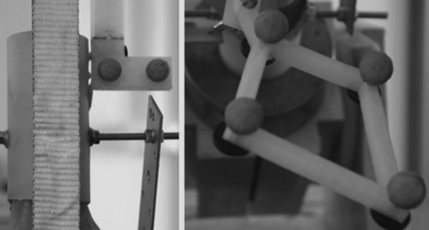
Navigation system. (A) L-shaped geometry frame for femur tracking; (B) asymmetrical rhomboids geometry for patellar tracking.
At first glance many still may wonder how simple 3D printed models can be having such a multi-tiered impact in medicine. By taking patient-specific data from MRIs or CTs and transferring it to a customized model, medical professionals are able to go much further than ever previously imagined. They are better able to diagnose and plot a course of treatment. From there they can also use the customized model to explain to the patient about their condition and outline following procedures. Surgeons can use the models to train for new and complex surgeries, navigate in the operating room by referring to the models, and then can allow them to work as training tools for medical students—a far cry from using a piece of fruit to practice on, or a cadaver.
With the use of 3D printed models, implants, and more, we’ve come a long way already from the one-size-fits-all mentality. With this new technology, we are moving into the era of true patient-specific treatment. 3D printed models also offer a look inside the human body almost like a snapshot that we can hold, examine, and use for research and insight into evaluating challenging conditions. How do you think these models will affect the future of similar research? Discuss in the 3D Printed Knee Part Models forum over at 3DPB.com.
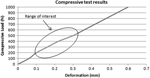
Compressive test results. The graph shows the compressive load as function of the deformation of the patella. In the figure, the range of interest for this study is circled.