
While the field may be advancing quickly, the process of creating 3D printed tissues and cells in the lab is a relatively slow and deliberate one. A person unfamiliar with the technology may picture doctors pulling out a 3D printer, turning it on, and – voila! – a 3D printed ear! In reality, 3D printed cells must be carefully cultivated and allowed to grow into viable tissue in a process that takes days at least. But a new development by researchers based at Australia’s University of Wollongong could speed that process up – with a tool that, in essence, is a biological 3Doodler.

The BioPen arose out of a collaboration between researchers at the UoW-based Australian Research Council Centre of Excellence for Electromaterials Science (ACES) and orthopedic surgeons at St. Vincent’s Hospital Melbourne. The device would allow surgeons to repair damaged bone and cartilage by “drawing” new cells directly onto bone in the middle of a surgical procedure. A team led by ACES Director Professor Gordon Wallace developed the pen, which was then transferred to St. Vincent for researchers to work on optimizing it for clinical trials.
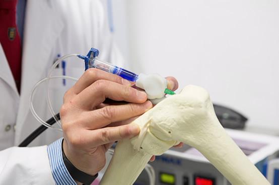
The technique could revolutionize how surgeons repair cartilage, in particular. For certain types of injuries, it’s difficult or impossible for surgeons to discern the exact shape of the area requiring an implant, making it extremely difficult to design an artificial cartilage implant before surgery. With the BioPen, surgeons could simply fill in the damaged area with the hydrogel solution.
“This type of treatment may be suitable for repairing acutely damaged bone and cartilage, for example from sporting or motor vehicle injuries,” said Professor Peter Choong, Director of Orthopaedics at St. Vincent’s. “Professor Wallace’s research team brings together the science of stem cells and polymer chemistry to help surgeons design and personalise solutions for reconstructing bone and joint defects in real time.”
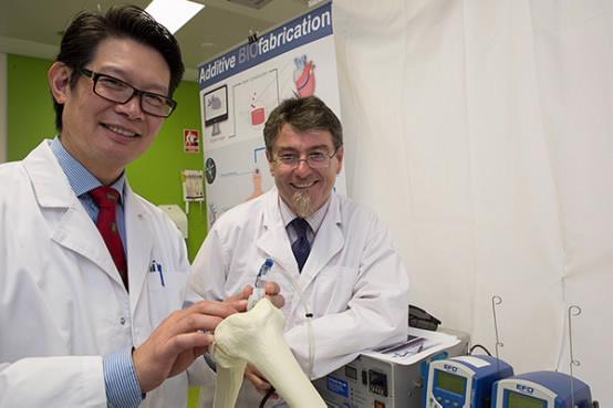
Professors Choong (L) and Wallace
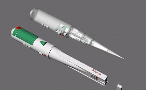
The cell solution could also be further customized by adding drugs to boost healing and regrowth. The BioPen prototype was 3D printed in medical grade plastic and titanium to be lightweight and easy to sterilize.
“The combination of materials science and next-generation fabrication technology is creating opportunities that can only be executed through effective collaborations such as this,” said Professor Wallace. “What’s more, advances in 3D printing are enabling further hardware innovations in a rapid manner.”
So far, cells produced by the BioPen have shown a survival rate of over 97%. The full research study has recently been published in the journal Biofabrication.
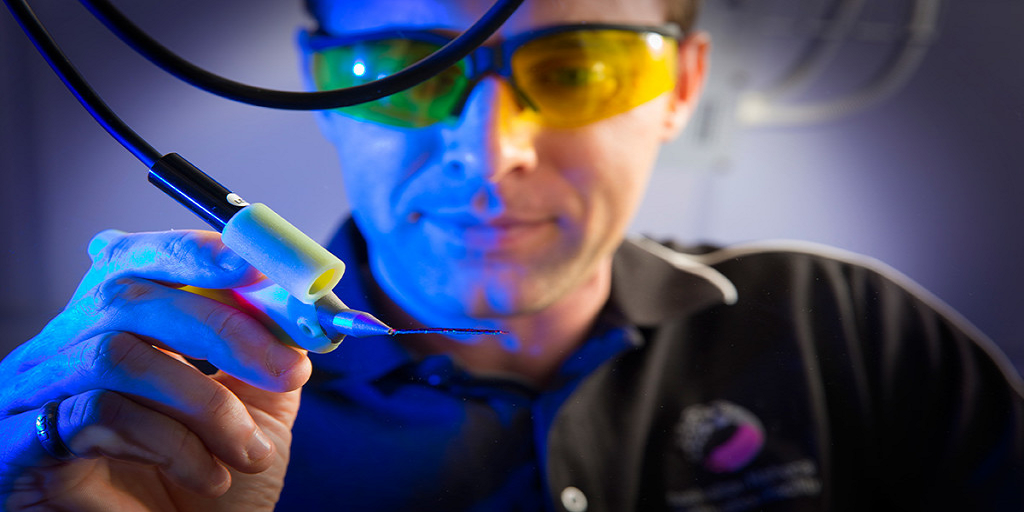
Below, Professor Choong demonstrates how the BioPen works. What do you think of the technology? Do you think it will catch on with surgeons? Discuss in the 3D Printing BioPen forum over at 3DPB.com.
