

[Image: Deidre Quinn-Gorham]
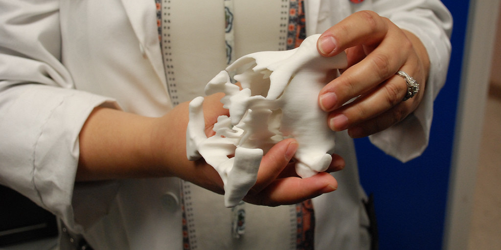
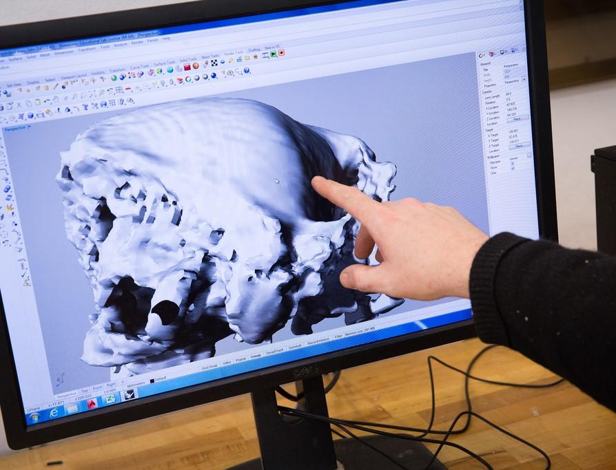
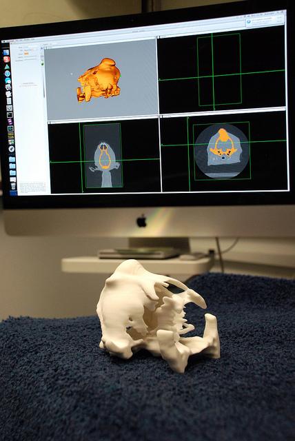
Evelyn Galban, a neurosurgeon who lectures at the University of Pennsylvania’s School of Veterinary Medicine, agrees. Dr. Galban recently became acquainted with a dog named Millie, who was brought in with a large tumor in her skull, one that was going to be difficult to treat – unless the vet could plan out the surgery ahead of time. Dr. Galban enlisted the help of the School of Design’s Fabrication Lab to turn Millie’s CAT scan data into 3D models, which were used to 3D print a replica of the dog’s skull. The gypsum model, which took about 6 hours to print, allowed Dr. Galban to closely examine the tumor, which protruded out from the top of Millie’s skull as well as inward, impacting her brain.
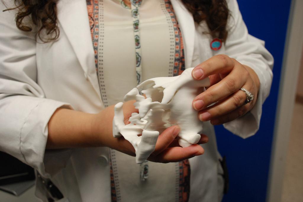
“It’s difficult to fully understand the malformation until we have it in our hands,” Dr. Galban said. “That usually doesn’t happen until we’re in surgery.”
We’ve seen this type of technology being used with more frequency in human surgeries, so it shouldn’t be surprising that it’s now starting to appear in veterinary procedures as well. Dr. Galban is working with PennDesign’s Stephen Smeltzer and Dennis Pierattini, along with veterinary residents Jon Wood and Leontine Benedicenti, to create 3D printed models of other dogs and cats suffering from deformities or injuries. Not only can these models help the individual animals, but they can also serve as valuable training aides for veterinary students.
“These objects have opened up to have applications in the real world, and that’s fascinating and enjoyable to see,” said Smeltzer. “Last week I had no idea that this was going to be happening, and now all of a sudden I have a vested interest in Millie.”
You can hear Dr. Galban describe the technology in greater detail here below. Are you surprised to hear that veterinarians are getting in on this technology too? Discuss in the 3D Printed Surgical Models for Animal Treatment & Training forum over at 3DPB.com.
https://youtu.be/dCC1i2Kv3bg
[Images: University of Pennsylvania]