One of the difficulties facing medical education is that the human body exists in three dimensions and requires study in three dimensions. This means that in order to appropriately understand human anatomy, pathologies, and development there is no substitute for hands-on experience with the human body. While that is still true there are at least new opportunities being presented through 3-D modeling and printing that can provide some of the experience previously only possible in working with cadavers.
To take this app to the next level, the team has turned to Indiegogo in an effort to raise $40,000 by September 20. This funding will be used to complete the cardiovascular simulation that they wish to include as a central component of the educational material. While there are certainly textbooks and illustrations already available, as well as plastic models demonstrating different aspects of embryological development, the team believes that the creation of this digital material is particularly important.
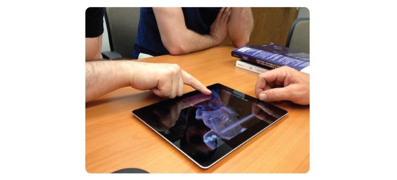
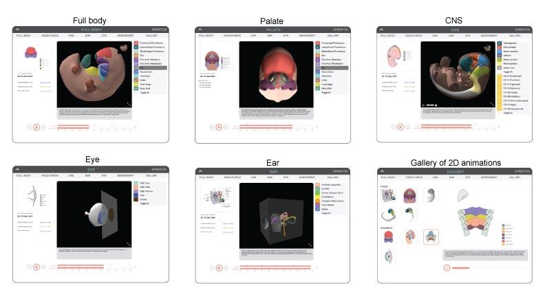
“Today’s students are digital natives and are comfortable using online and digital assets in their study… Previous work has shown that the presentation of embryological structures as 3-D models rather than using 2-D illustrations enhances a student’s understanding of the morphogenesis of complex tissues and organs. A student can grasp the complexities of facial development or heart development much more quickly and with deeper understanding through the use of the 3-D model rather than 2-D illustrations.”
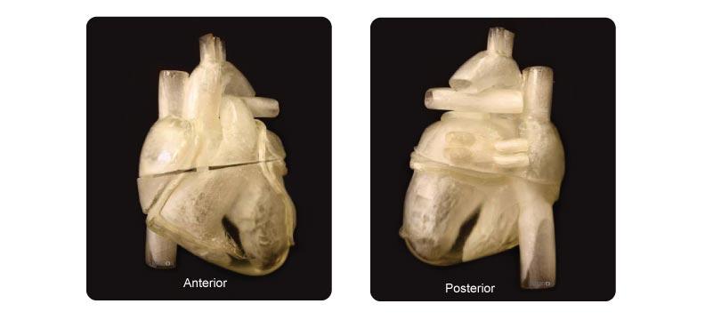
“For example, in the heart embryonic simulation, the student learns how the primitive part two subdivides into the four chambers of the fetal and adult heart. The student is also presented with material to understand the congenital heart anomalies that result from the failure to complete formation of these chambers. Once a student has completed the digital material and assessment in our application, they will be presented with the same heart model from the application, but as a physical 3-D printed model, that depicts either normal development formation of a congenital anomaly.”
With congenital anomalies, commonly referred to as first effects, affecting approximately one in every 33 infants and resulting in hundreds of thousands of newborn deaths every year, it is vital that medical and dental students have as many opportunities as possible to fully understand the anomalies, their implications, and possible interventions. The creation of this app and the accompanying physical models will provide students with much greater access to the resources that they need as they prepare for practice.
3D printing as a means for medical instruction is becoming more and more common these days. Let us know your thoughts on this creative use of 3D printing in the 3D Printed human embryo forum thread on 3DPB.com. Check out the video below for further details on this Indiegogo campaign.