
3D Printed Personalized Mold Contributes to Advances in Prostate Cancer Diagnoses

Advances in imaging techniques are changing the way in which prostate cancer can be detected and treated. However, these techniques have yet to be perfected and there is room for a great deal of improvement. To this end, a team of researchers from the University of California Los Angeles and the David Geffen School of Medicine, also in Los Angeles, worked together to pinpoint the areas of current imaging techniques that required modification and improvement.
 In their most recent paper, printed in the American Journal of Clinical and Experimental Urology, the team described a low cost, 3D printed, patient specific mold, designed to support a MR-histology correlation process, in which the data gathered on the prostate through an MRI was compared with the tissue that was removed during a radical prostatectomy. By examining the actual presence and extent of cancer in the removed tissue with the indications presented through the MRI data, the team was able to pinpoint areas of the MR process that require refinement.
 For this particular study, a 54-year-old patient who was to undergo a radical prostatectomy gave permission to use the imaging and tissue that would allow the team to study the veracity of the image data. The MRI he had been given, had indicated a high level of suspicion for cancer that was organ confined. Therefore, he had decided to undergo treatment that included a radical robotic prostatectomy, designed to remove the prostrate gland and some of the surrounding tissue.
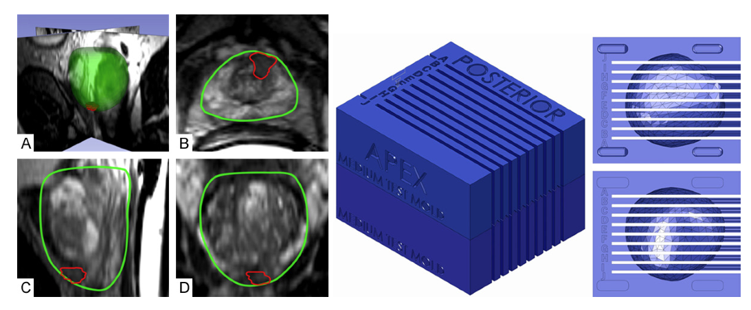
The prostate cavity mold is designed to hold the excised tissue in the position necessary for an exact comparison with the MRI data. The form of the interior cavity of the mold is created from by using the 3D imaging software, Profuse, developed by Eigen, that generates true, navigable, and quantifiable 3D images from ultrasound. The “slice” images, or segmentations, that were used to build up the 3D model of the prostrate were then imported into SolidWorks in order to generate the mold cavity that would perfectly match the tissue surface.
The mold itself was made from polylactic acid plastic (PLA) and printed on Makerbot’s Replicator 2. It was manufactured in two halves with a central cavity in which the tissue is placed according to the position in the MRI. It also has a series of slits, in this case placed 4.5 mm apart, in order to correspond to the MR image planes. The printing of the mold was completed prior to the surgery and was ready in the grossing room, the area where pathology specimens from the operating room is taken for pathological review and analysis, when the operation commenced.
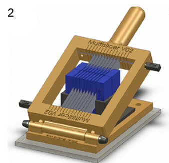 When the excised tissue specimen was delivered to the grossing room, a 3D scan was produced using Makerbot’s Digitizer 3D scanner. After recording this data, the mold was placed in a custom made slicing apparatus that pushed the blades through the slits left in the mold, creating 10 segments, each corresponding with a segment image created by the MRI.
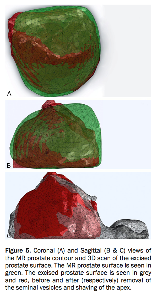
The team then worked to carefully study the data that had been available prior to the surgery through the MRI, with the reality of the condition of the excised tissue in a process known as MR-histology correlation. In comparing the data, the team discovered that while the MRI had accurately predicted the presence of the cancerous regions, it had produced data that indicated a significantly smaller lesion volume and extent.
The MRI reads the presence of cancer using the report generated by pulsing signals, T1 and T2 signals. The researchers noted that the cancer-positive regions discovered through the histology that had not been made visible through the MRI were observed to have low T2 signal responsiveness. This data indicates, therefore, the need for a refinement, to improve the MR sensitivity in order to increase the predictive capabilities of the images created.
By continuing to refine the images produced by an MRI, not only is the likelihood of greater detection accuracy increased, it also provides the surgeons with a better roadmap of what they are likely to encounter as they perform the procedure. In addition, to improvement in the possibilities for detection is also a lower rate of false positives that may lead to unnecessary surgeries, further testing, or other treatments.
The difficulties of studying the relationship between the imaged data and the actual tissue has been present, largely due to the deformation of the shape of the tissue once it has been removed from its site in the body. The 3D printed mold is designed to mitigate exactly this issue and allow for direct examination of the tissue in its in situ shape.
This, like so many other changes and advances in medical technology that involve 3D printing, builds upon the absolute personalization that is allowed by the technology. A great deal more research is necessary in order to confirm the findings gathered during this study, but one thing is for certain, 3D printing is contributing to addressing medical issues in an increasingly wider variety of ways, while allowing investigations to begin that previously had been impossible.
Discuss this incredible use of 3D printing in the Prostate Cancer Diagnoses forum thread on 3DPB.com
Subscribe to Our Email Newsletter
Stay up-to-date on all the latest news from the 3D printing industry and receive information and offers from third party vendors.
You May Also Like
Precision at the Microscale: UK Researchers Advance Medical Devices with BMF’s 3D Printing Tech
University of Nottingham researchers are using Boston Micro Fabrication‘s (BMF) 3D printing technology to develop medical devices that improve compatibility with human tissue. Funded by a UK grant, this project...
3D Printing Webinar and Event Roundup: April 21, 2024
It’s another busy week of webinars and events, starting with Hannover Messe in Germany and continuing with Metalcasting Congress, Chinaplas, TechBlick’s Innovation Festival, and more. Stratasys continues its advanced training...
3D Printing Webinar and Event Roundup: March 17, 2024
It’s another busy week of webinars and events, including SALMED 2024 and AM Forum in Berlin. Stratasys continues its in-person training and is offering two webinars, ASTM is holding a...
3D Printed Micro Antenna is 15% Smaller and 6X Lighter
Horizon Microtechnologies has achieved success in creating a high-frequency D-Band horn antenna through micro 3D printing. However, this achievement did not rely solely on 3D printing; it involved a combination...


