Scientists Use 3D Printed Models to Further Congenital Heart Disease Studies

In the recently published ‘Accurate Congenital Heart Disease Model Generation for 3D Printing,’ researchers explore 3D printing for diagnosis, treatment, and planning in congenital heart disease (CHD) patients. CHD usually presents itself at birth and can be difficult to analyze, even with 3D medical images—and despite many different studies by scientists around the world. The researchers note that 3D printing has been ‘widely adopted’ in clinical settings lately, offering for improvements in the following:
- Clinical decision making
- Interventional planning
- Communication between physicians and patients
- Improving medical education
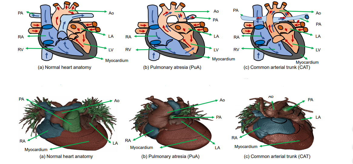
(Top) Examples of large structure variations in CHD. In normal heart anatomy (a), PA is connected to RV. However, in pulmonary atresia (b), PA is rather small and connected to descending Ao. In common arterial trunk (c), Ao is connected to both RV and LV, and PA is connected to Ao. (Bottom) Pulmonary atresia and common arterial trunk examples in our dataset, with large variations from normal heart anatomy.
In the dataset for this research, combining deep learning and graph matching for whole heart and great vessel segmentation in CHD, patients ranged in age from one month to 21 years old—while most were from one month to two years old. Out of 16 cases, the study covers 14 types of CHD to include the most common 8, which are atrial septal defect (ASD), atrio-ventricular septal defect (AVSD), patent ductus arteriosus (PDA), pulmonary stenosis (PS), ventricular septal defect (VSD), co-arctation (CA), Tetrology of Fallot (ToF), and transposition of great arteries (TGA).

Overview of the proposed framework combining deep learning and graph matching for whole heart and great vessel segmentation in CHD.
The researchers explain that there has already been a prolific amount of research using the multi-modality whole heart segmentation method, with ‘state-of-the-art performance’ found in combining 3D U-net for segmentation and a simple convolutional neural network for label position prediction. Another technique involves using the basic simple convolutional neural network for label position prediction, while another handles blood pool and myocardium in blood pool segmentation.
“Considering the significant variations in heart structures and great vessel connections in CHD, almost all the existing methods cannot effectively perform whole heart and great vessels segmentation in CHD,” state the researchers.
Motivated instead by the promise of graph matching, they used deep learning and graph matching, collecting 68 CT images overall, with an 11.9 percent higher Dice score. The framework consisted of:
- Region of interest cropping
- Chambers and myocardium segmentation
- Blood pool segmentation
- Chambers and myocardium refinement
- Graph matching
Segmentation results were printed on a Sailner J501Pro for evaluation—a process that took the researchers about three to four hours. The researchers assessed the 3D printed model as correct, and with clear shape and connections, with minor refinements necessary such as thin coronary vessels.
“We also printed out part of the segmentation results with minor manual refinement and showed that it can be applied to clinic use,” concluded the researchers.
3D printed models have proven to be more than just helpful in many different areas of the medical realm, especially as they are able to offer so much in terms of education not only for students but also for patients and their families, as well as helping in the preoperative stages—and during the actual operation too with surgical planning models.
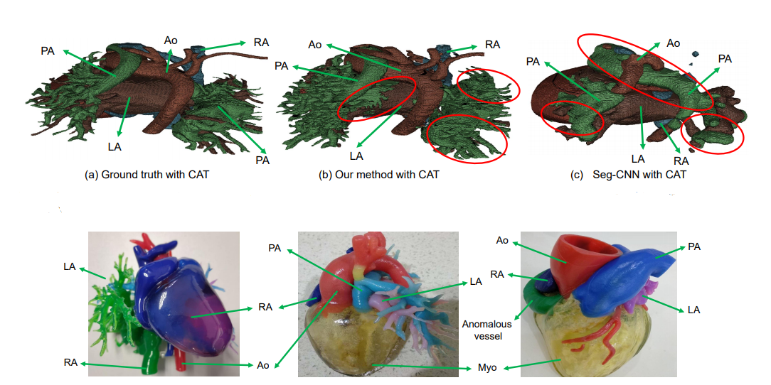
(Top) Visualized comparison between the state-of-the-art method Seg-CNN [12] and our method. The differences from the ground truth are highlighted by the red circles. (Bottom) Examples of 3D printing models using our method with some minor manual refinement.
Subscribe to Our Email Newsletter
Stay up-to-date on all the latest news from the 3D printing industry and receive information and offers from third party vendors.
You May Also Like
Air Force Awards Fortius Metals $1.25M to Qualify 3D Printing Wire for Hypersonic Applications
AFWERX, part of the US Air Force Research Laboratory (AFRL), awarded a Direct-to-Phase II Small Business Innovation Research (SBIR) contract worth $1.25 million to Colorado’s Fortius Metals, to accelerate qualification...
US Air Force Awards JuggerBot $4M for Large-format Hybrid 3D Printing
Large-format 3D printer manufacturer JuggerBot has received a $4 million grant to develop a large format 3D printer, courtesy of the Under Secretary of Defense, Research and Engineering Manufacturing Technology...
Where Have All AM’s Unicorns Gone?
In the rapidly evolving world of 3D printing, startups valued at over a billion dollars, known as unicorns, once seemed as fantastical as the mythical creatures themselves. While a few...
How My Childhood Fascination with Planes Led to Investing in 3D Printing
My fascination with aerospace started young, and I started studying planes–identifying them in the sky and learning everything I could about how they work. Fast forward to my first week...